

Colonoscopy quality indicators help clinicians and endoscopy units assess whether an examination was complete, careful, safe and useful for the patient. A colonoscopy is more than reaching the end of the colon: quality also depends on bowel cleansing, inspection, recognition and removal of lesions, documentation, communication and appropriate follow-up.
For gastroenterologists and endoscopy teams, measuring performance turns these principles into opportunities for improvement. This guide explains seven practical areas to monitor, how common indicators are defined, and how teams can use results constructively. Numerical targets cited from US guidance are identified as such; SGEI readers should interpret them alongside applicable Indian guidance, local protocols and patient context.
What do colonoscopy quality indicators measure?
Quality indicators are measurable aspects of care that can be reviewed at the endoscopist or unit level. They cover the structure and process of an examination as well as outcomes and patient experience. One strong measure cannot prove that every part of a colonoscopy was high quality. For example, reaching the caecum does not guarantee careful mucosal inspection, and detecting lesions does not establish that they were completely and safely removed.
The 2024 ASGE/ACG update identifies priority colonoscopy indicators including adenoma detection rate (ADR), sessile serrated lesion detection rate (SSLDR), bowel preparation adequacy, use of recommended screening and surveillance intervals, and caecal intubation. These are useful reference measures, not a substitute for locally agreed standards. Read the ASGE/ACG 2024 quality indicator FAQ.
A quick view of commonly used US reference targets
| Indicator | 2024 US reference target | What it helps assess |
|---|---|---|
| Bowel preparation adequacy | ≥90% | Whether preparation supports standard interval assignment |
| Caecal intubation rate | ≥95% | Completion and landmark documentation |
| Adenoma detection rate | ≥35%* | Detection of conventional adenomas |
| Sessile serrated lesion detection rate | ≥6%* | Detection of serrated precancerous lesions |
| Mean withdrawal time | ≥8 minutes* | Time devoted to systematic inspection |
Thresholds apply to defined populations and denominators in the US guideline. Verify eligibility, exclusions and current local recommendations before comparing performance. The measures complement one another; no single target captures the entire quality of an examination.
1. Bowel preparation adequacy
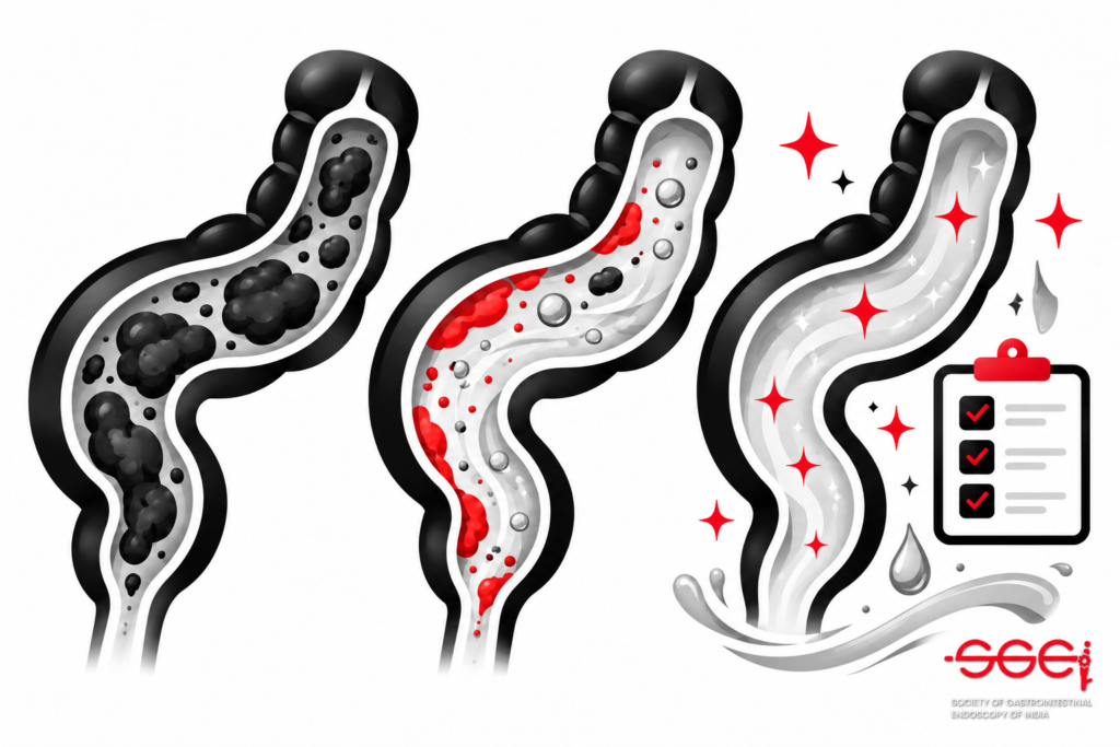
A clean colon allows the endoscopist to inspect the mucosa reliably. Residual stool or opaque liquid can hide lesions, prolong the procedure, impair caecal intubation and make follow-up planning uncertain. Preparation quality is therefore a shared responsibility: patient instructions, regimen selection, timing, nursing support and endoscopic cleaning all contribute.
Document preparation quality using a consistent, validated scale after washing and suctioning. The Boston Bowel Preparation Scale (BBPS), for example, scores the right, transverse and left colon separately. The 2024 US ASGE/ACG indicator defines adequate preparation in relation to whether standard screening or surveillance intervals can be assigned; it sets a target of at least 90% adequacy for outpatients. Units should specify their local definition and include cancellations for presumed inadequate preparation when auditing the process.
When preparation is inadequate, explain the limitation clearly, record the segment affected and arrange a repeat examination at an interval appropriate to the indication and current guidance. The US Multi-Society Task Force bowel preparation recommendations offer practical guidance on improving preparation and follow-up. Adapt recommendations to local practice and patient needs.
2. Caecal intubation and complete examination
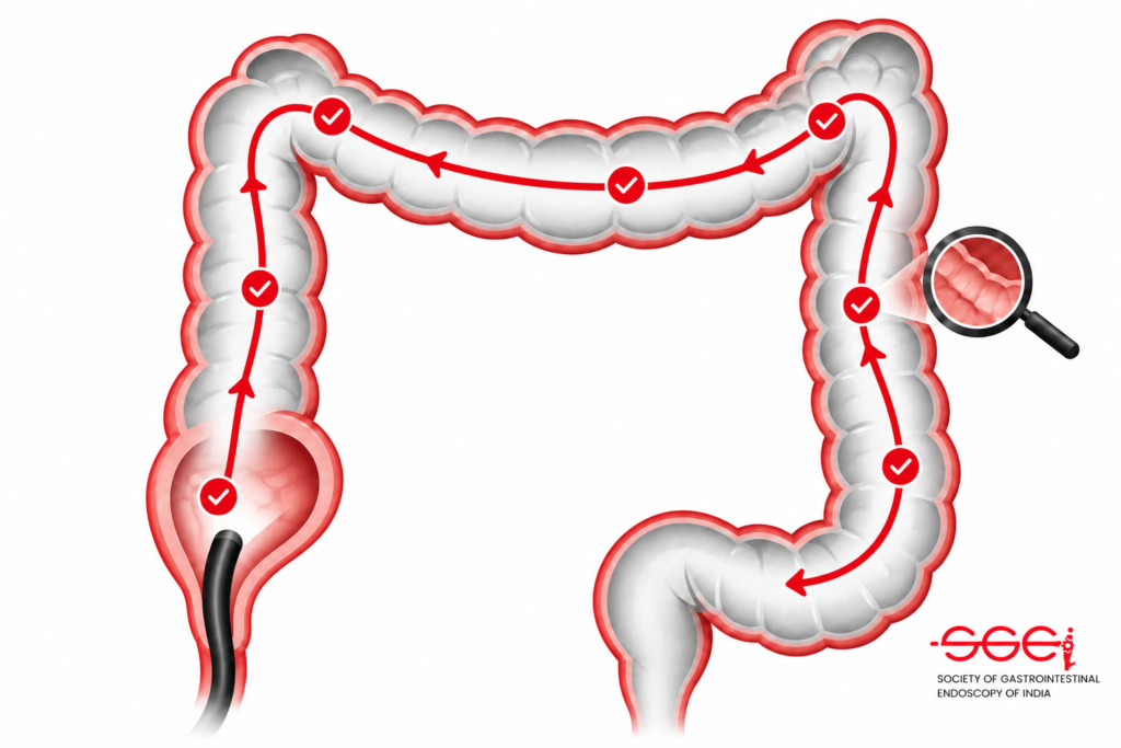
A screening or diagnostic colonoscopy should reach the caecum when clinically appropriate. Caecal intubation rate (CIR) is the proportion of eligible procedures in which the caecum is reached and adequately documented. The 2024 US ASGE/ACG target is at least 95%, with photographic evidence of landmarks such as the appendiceal orifice and ileocaecal valve. Exclusions and denominators should follow the chosen standard and be applied consistently.
A high CIR is not simply a race to the endpoint. Safe insertion, patient comfort, loop management, adequate sedation planning and careful confirmation of landmarks matter. If the examination is incomplete, document why, what was visualised and the plan to complete colonic assessment whether by repeat colonoscopy with an appropriate strategy or another suitable test. Review incomplete cases for patterns such as preparation, anatomy, discomfort, equipment or training needs.

3. Adenoma detection rate
ADR is the proportion of eligible patients undergoing colonoscopy in whom at least one conventional adenoma is detected and confirmed by pathology. It is an important measure because missed precancerous lesions can contribute to post-colonoscopy colorectal cancer. The denominator must be defined carefully: patient age, indication, positive stool-based or imaging screening tests, inflammatory bowel disease, hereditary syndromes and procedures performed for known lesions can affect inclusion under a given guideline.
The 2024 US ASGE/ACG update sets an overall ADR target of at least 35% for specified screening, surveillance and diagnostic examinations in people aged 45 years or older, with sex-specific targets also provided. These are US benchmarks and should not automatically be treated as universal thresholds for every Indian population, setting or case mix. Compare like with like, use accurate pathology linkage and review trends over time rather than reacting to a small sample.
ADR is a rate, not a complete description of technique or patient benefit. It can be affected by case mix and documentation. Teams can support careful detection with adequate preparation, sufficient inspection, appropriate use of position changes and washing, and review of endoscopy images and pathology feedback. Use audit results for learning and improvement, not as a standalone ranking.
4. Sessile serrated lesion detection
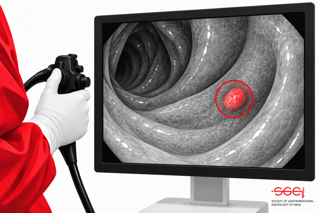
Sessile serrated lesions (SSLs) can be subtle and are often flat, pale or covered by mucus. Their recognition and management form an important part of a complete inspection. The ASGE/ACG 2024 update added SSL detection rate as a priority outcome measure and gives a US performance threshold of at least 6% in the defined population.
To measure SSLDR, units need reliable endoscopy documentation linked to pathology, consistent terminology and clear denominator rules. Pathology reports may use older terms; agree how these map to the measure and align reporting language with current practice. A lower rate can prompt a structured review of preparation, withdrawal technique, image quality and pathology concordance. As with ADR, interpret the result in light of case mix and local standards.
5. Withdrawal time and careful inspection
Withdrawal is the phase in which the endoscopist systematically inspects the mucosa while withdrawing the scope. Adequate time supports a deliberate examination, but time alone does not guarantee quality. The ASGE/ACG 2024 guidance uses a mean withdrawal-time threshold of at least eight minutes for normal examinations in the specified population, with defined exclusions. This is a measure to interpret alongside detection outcomes, not a target to meet by waiting without careful inspection.
A consistent withdrawal approach includes cleaning residual material, distending the lumen, inspecting behind folds where feasible and documenting important findings. Endoscopy units can review withdrawal times alongside ADR, SSLDR and preparation adequacy to identify where further training or workflow changes may help. The aim is a careful, reproducible examination that is appropriate to the patient and indication.
6. Polypectomy quality and safety
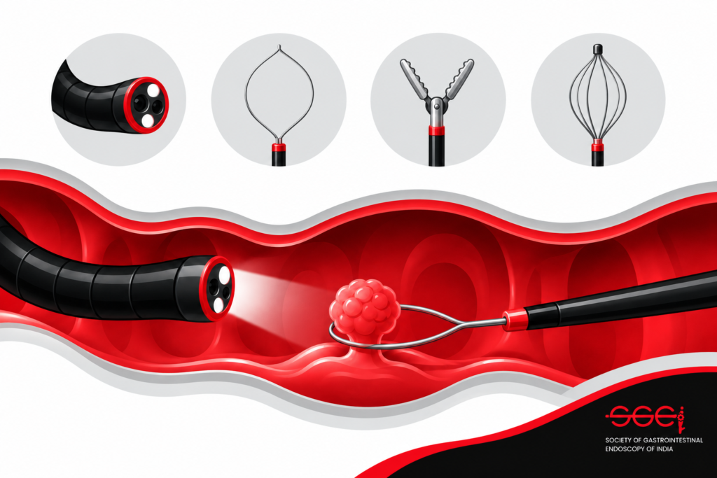
Finding a lesion is only one part of care. The endoscopist must assess its features, choose an appropriate management strategy, resect safely when indicated, retrieve tissue where appropriate and document completeness. Technique depends on lesion size, morphology, location, patient factors and available expertise. Complex lesions may require referral to an appropriately experienced endoscopist rather than an attempted resection outside the clinician’s competence.
Track adverse events and unplanned care according to local policy, including bleeding, perforation, sedation-related events and post-procedure presentations. Consistent definitions, adequate follow-up and fair attribution are essential for useful audits. Review complications in a learning-focused process that looks at the patient, procedure, equipment and system factors. A low event count in a small sample does not by itself establish safety.
7. Documentation, follow-up and patient experience
A useful report makes the examination understandable to the next clinician and to the patient. Record indication, bowel preparation, extent reached and landmarks, findings and their locations, interventions, specimens, complications, limitations and the recommended follow-up plan. Link pathology results to the endoscopy record and ensure responsibility for communicating results is clear.
Surveillance recommendations should be based on the complete examination, pathology, personal and family history, relevant guidelines and patient factors. Avoid recommending an interval that cannot be supported by examination quality or the findings. The 2024 US ASGE/ACG guidance includes adherence to recommended screening and surveillance intervals as a priority indicator. SGEI members can also explore SGEI’s education and academic activities to stay connected with endoscopy learning in India.
Patient experience is another meaningful dimension. Clear preparation instructions, respectful communication, comfort assessment, privacy, timely explanations and accessible discharge guidance can improve the care journey. Collect feedback in a consistent way and use it alongside clinical measures; satisfaction scores should not replace measures of examination quality or safety.
How can an endoscopy unit improve quality?
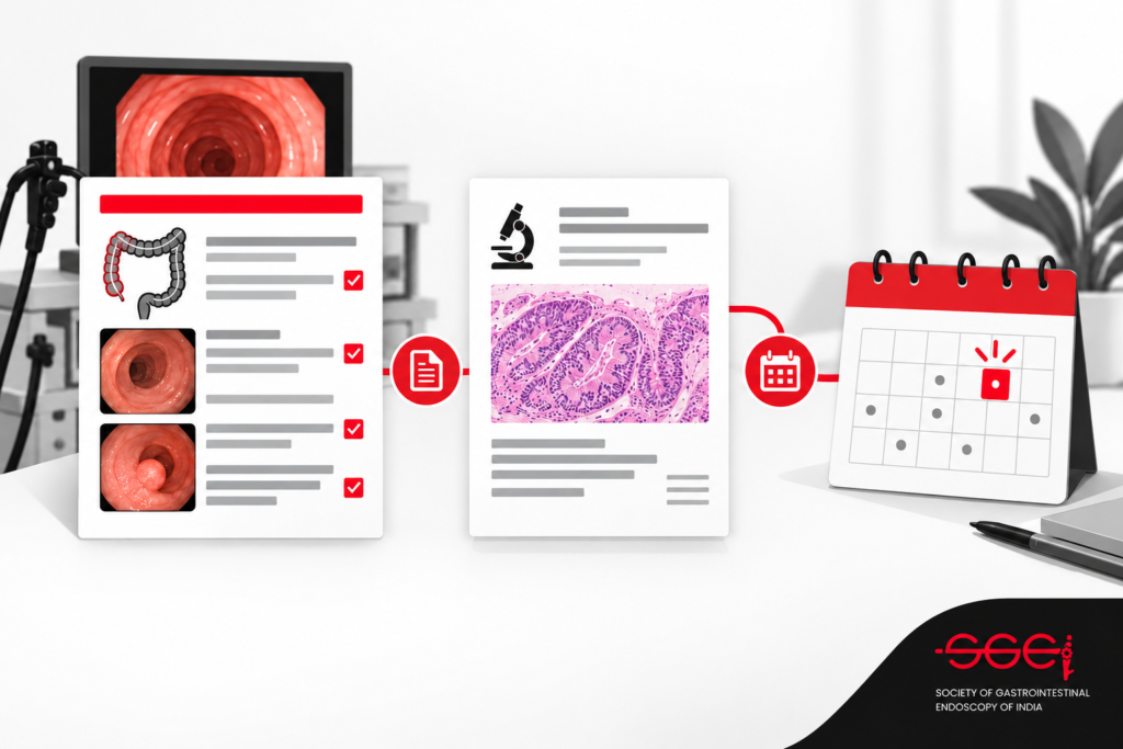
Start with a small, reliable audit. Agree on indicator definitions, eligible cases, exclusions, data sources and review frequency before comparing results. Automate data capture where possible, check a sample of records for accuracy and link pathology and follow-up data. Report unit-level patterns and provide individual feedback confidentially and constructively.
When a measure falls below the locally selected threshold, look for a process that can be changed. Examples include revising bowel-preparation instructions, improving pre-procedure assessment, standardising landmark photographs, reviewing inspection technique or clarifying who communicates pathology results. Set a specific improvement action, measure again and share what the team learns. Benchmarks should guide investigation, not encourage rushed procedures, unnecessary interventions or avoidance of complex patients.
A practical dashboard can display monthly or quarterly trends for each agreed indicator, with the denominator and exclusions shown beside the result. Stratifying results by indication or site may reveal a workflow issue that an overall average hides. Protect patient confidentiality, validate automated reports and involve endoscopists, nurses, administrators and pathology teams in interpreting results. Quality improvement works best when the people who do the work help choose a change that is realistic in their setting.
Conclusion: Make colonoscopy quality measurable
High-quality colonoscopy combines adequate preparation, complete and safe examination, careful lesion detection, appropriate resection, clear documentation and suitable follow-up. Colonoscopy quality indicators help endoscopists and units understand how reliably these elements are delivered and where targeted improvement may benefit patients.
Use current professional guidance, define measures consistently and interpret results in context. For SGEI readers, international targets are useful reference points; local standards and patient populations should shape implementation. A thoughtful audit paired with practical team learning can turn measurement into better, more consistent endoscopy care.
Frequently asked questions
What are the most important colonoscopy quality indicators?
Common priority indicators include bowel preparation adequacy, caecal intubation, adenoma detection, sessile serrated lesion detection and appropriate screening or surveillance recommendations. The exact set and thresholds depend on the guideline used.
What is a good adenoma detection rate?
The 2024 US ASGE/ACG guideline sets a target of at least 35% for a defined group of examinations, with sex-specific thresholds. It is a US benchmark; interpret it using the full guideline definitions and applicable local standards.
Does a longer withdrawal time always mean a better colonoscopy?
No. Withdrawal time is a supportive process measure. It should accompany careful inspection and outcome measures such as lesion detection, with exclusions and definitions applied consistently.
What should happen after inadequate bowel preparation?
Document the limitation, explain its effect on examination reliability and arrange repeat or alternative assessment at an interval suited to the indication and current guidance.