Table of Contents
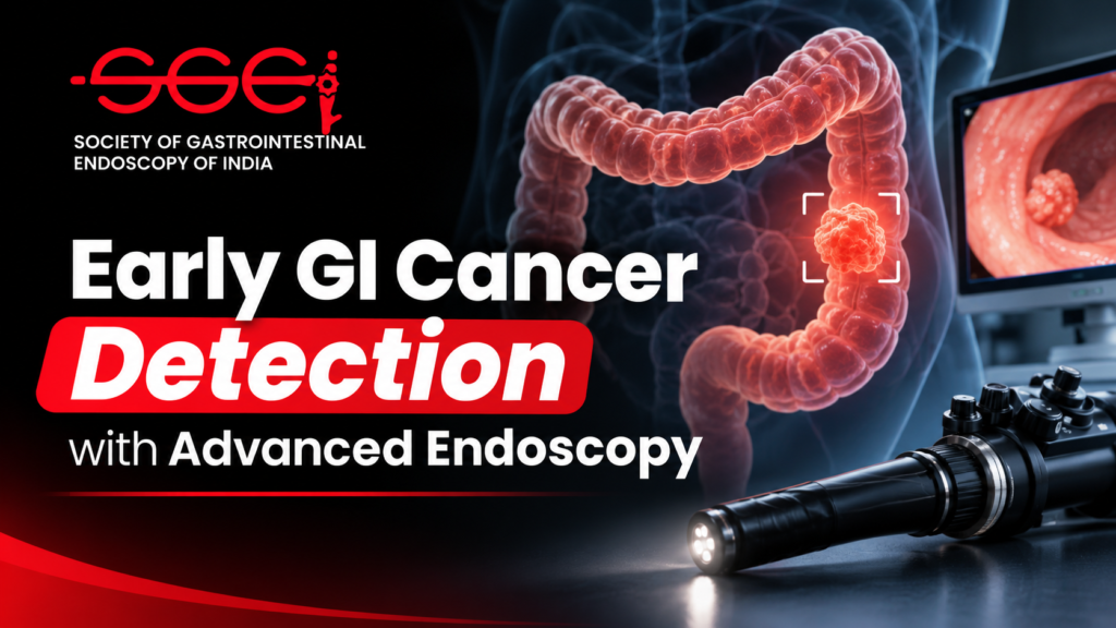
Early GI cancer detection can change the decisions available to patients and clinicians. Many gastrointestinal cancers develop through visible precancerous changes or early disease, creating opportunities for recognition, biopsy, surveillance or endoscopic treatment before disease becomes more advanced. Endoscopy is uniquely useful because it allows a clinician to inspect the lining directly, target suspicious tissue and, in selected cases, remove early lesions. The European Society of Gastrointestinal Endoscopy (ESGE) describes endoscopy as an important tool for detecting GI cancers and precursor lesions, while emphasizing that screening recommendations depend on cancer type and risk group in its position statement on early diagnosis and prevention.
The challenge is that early lesions may be flat, small, pale, irregular, or visually similar to surrounding tissue. A conventional white-light examination can miss subtle borders or changes in surface and blood-vessel patterns, particularly when visibility is poor or inspection is hurried. Advanced endoscopic imaging helps make these features easier to see and interpret. It supports early GI cancer detection by adding contrast and detail to a careful examination; it does not replace clinical judgement, pathology or an appropriate screening strategy.
What advanced endoscopic imaging adds
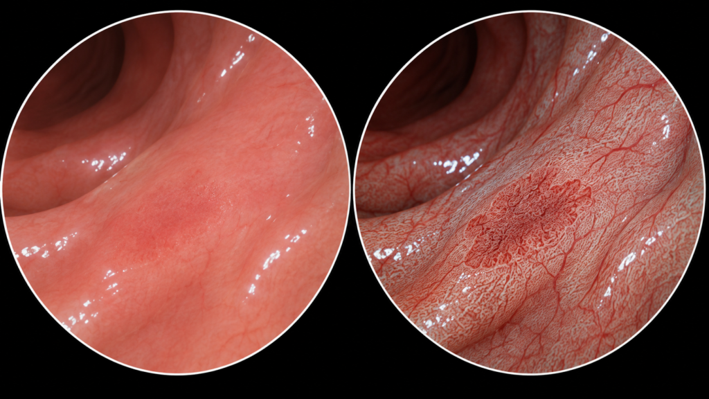
High-definition white-light endoscopy remains the foundation. The endoscopist first cleans the mucosa, obtains a stable view, inspects the full relevant area and notes abnormalities in context. Image-enhanced endoscopy (IEE) adds optical or digital contrast that can make mucosal relief, surface architecture and microvascular patterns more conspicuous. Magnification can then enlarge a suspicious area for a closer assessment.
These views can help answer practical questions: Is this area different from the surrounding mucosa? Are its borders clear? Does the surface or vascular pattern look regular or disrupted? Is the appearance consistent with a benign change, dysplasia or possible invasion? In selected settings, a trained endoscopist can use validated optical classifications to estimate histology and guide targeted biopsy or treatment planning. ESGE’s optical-diagnosis curriculum highlights these skills for Barrett’s oesophagus, squamous oesophageal lesions, early gastric cancer, colorectal lesions and inflammatory bowel disease. SGEI’s SITAR training and research initiatives and Young Endoscopist UpSkilling (YES) programme are relevant places to check for professional learning opportunities; confirm current availability and eligibility on the official pages.
Advanced endoscopic imaging can focus attention, help delineate lesion margins and support characterization. For early GI cancer detection, it informs recognition, documentation and sampling but optical appearance alone does not establish cancer; pathology and clinical judgement remain essential.
Core imaging technologies used for early GI cancer detection
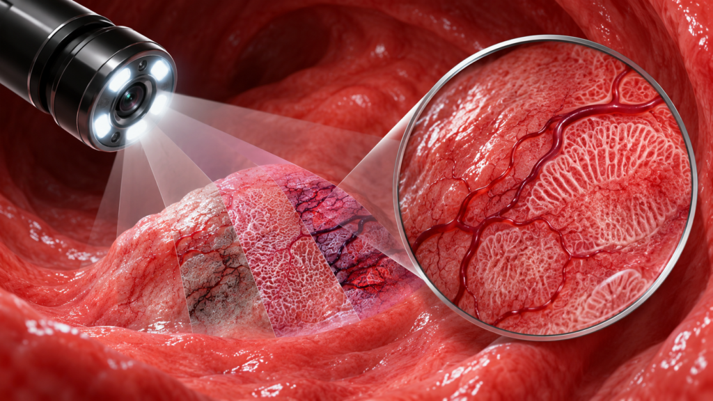
Several imaging approaches can support early GI cancer detection. Availability differs by endoscopy platform, and no single mode is best for every lesion or anatomical site.
Narrow-band imaging and virtual chromoendoscopy
Narrow-band imaging (NBI) uses selected wavelengths of light to emphasize superficial blood vessels and mucosal patterns. Other virtual chromoendoscopy systems use digital processing or alternative light spectra to enhance contrast. These modes can be activated during endoscopy without applying dye. In the stomach, the 2025 ESGE/EHMSG/ESP guideline reports evidence supporting virtual chromoendoscopy, including NBI, blue-laser imaging (BLI) and linked-color imaging (LCI), for detecting early lesions and precancerous conditions. It recommends using virtual chromoendoscopy to guide biopsies of suspicious areas.
Dye-based chromoendoscopy
Dye-based techniques apply a contrast agent to the mucosa. Depending on the indication, dyes can accentuate surface topography or highlight areas that differ in their response to staining. This can be useful for mapping a lesion’s edges or examining selected high-risk areas. The choice of dye and protocol should reflect the suspected condition, local expertise and current guidance.
Magnification and optical diagnosis
Optical magnification lets the endoscopist inspect surface and microvascular patterns at higher detail, often together with NBI or another contrast mode. Validated classification systems help translate what is seen into a structured description. This can support decisions about lesion characterization and referral for endoscopic resection, but performance depends on training and experience. Optical diagnosis complements histopathology; it does not justify skipping tissue assessment when biopsy or resection pathology is indicated.
Confocal endomicroscopy and other advanced methods
Confocal laser endomicroscopy and related technologies can provide microscopic-level images in selected specialist settings. They may help characterize tissue in real time, but access, training requirements, cost and evidence vary. These tools are not required for every routine examination. The core of early GI cancer detection remains a high-quality examination with a suitable imaging method, adequate documentation and a clear plan for abnormal findings.
Where early GI cancer detection can benefit
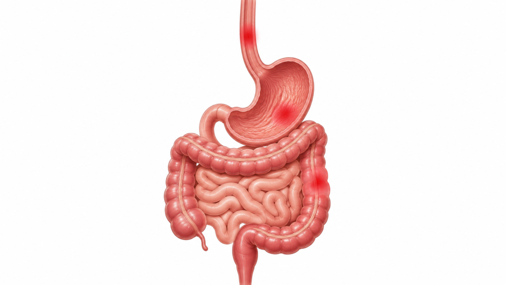
Advanced endoscopic imaging is used across the gastrointestinal tract, with specific techniques chosen for the disease, patient risk and clinical question.
Oesophagus
In Barrett’s oesophagus surveillance, careful inspection and chromoendoscopy can help identify focal areas that merit targeted assessment. In people at elevated risk for squamous neoplasia, image enhancement may make subtle mucosal or vascular changes more visible. Screening decisions should follow appropriate risk-based guidance; these techniques do not mean that every person with reflux needs endoscopic cancer screening. ESGE’s position statement recommends considering oesophageal screening in selected high-risk groups rather than treating it as universal population screening.
Stomach
Early gastric cancer and precancerous gastric conditions can be subtle. A systematic examination with good mucosal visibility, adequate cleansing and careful inspection is essential. Virtual chromoendoscopy can help characterize suspicious areas and direct targeted biopsies. The 2025 ESGE guideline also notes that training in gastric neoplasia detection improves detection rates, reinforcing that the imaging mode and the operator’s technique work together.
Colon and rectum
For colorectal lesions, enhanced contrast can help distinguish neoplastic from non-neoplastic patterns and define lesion features. Validated classifications support optical diagnosis in carefully selected contexts. ESGE guidance on advanced imaging for colorectal neoplasia allows optical diagnosis of diminutive polyps only under controlled conditions, with trained endoscopists, validated scales, adequate documentation and audit. These limits matter: a promising image should not become an unsupported treatment decision.
A systematic workflow for early GI cancer detection
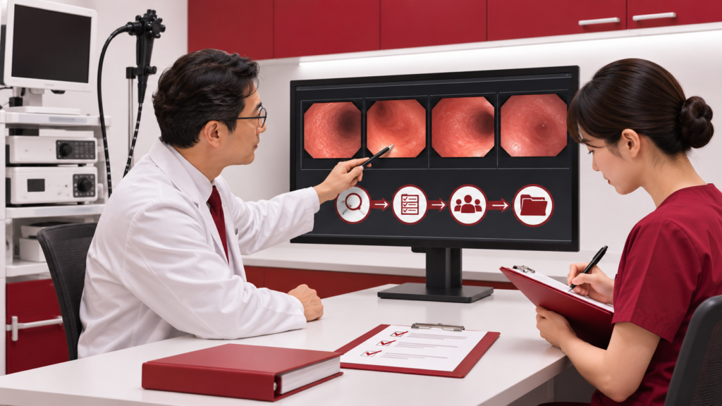
Advanced endoscopic imaging works best as part of a consistent workflow. Before the procedure, establish the indication, relevant risk factors, prior findings and the examination plan. Explain the procedure and obtain consent according to local practice. In high-risk surveillance, review the condition-specific protocol and ensure the required equipment is available.
During the examination, start with a careful high-definition white-light assessment. Clean mucus, bubbles and debris; distend the lumen as appropriate; and inspect systematically rather than relying on a rapid pass. Use image enhancement when it can clarify a suspicious area or when a guideline-supported protocol calls for it. Compare the abnormality with nearby mucosa and use magnification or a validated classification when available and within the endoscopist’s competence.
If a lesion is found, document its location, size, morphology and visual pattern. Capture images that show both the lesion and its relationship to surrounding anatomy. The 2025 ESGE gastric guideline calls for suspicious lesions to be properly described and photodocumented, followed by targeted biopsies. The exact sampling and treatment plan should reflect the site, appearance, local guidelines and specialist judgement.
After the procedure, connect the endoscopic impression with pathology and communicate a clear next step. That may be routine follow-up, targeted surveillance, endoscopic resection by an appropriately skilled team, multidisciplinary review or further staging. The workflow closes only when someone is responsible for results and the patient understands what happens next. This disciplined sequence is a practical foundation for better early GI cancer detection, regardless of the specific brand of imaging system.
At unit level, early GI cancer detection can be strengthened by agreeing on a minimum image set and structured lesion descriptions. Review whether key views were captured, whether pathology matched the endoscopic impression, and whether follow-up was completed. Use these findings to guide team learning; do not turn them into simplistic rankings, since case mix, available equipment and training access differ among centres. For a related framework for measuring procedure quality, see SGEI’s colonoscopy quality indicators guide.
The role of AI in early GI cancer detection
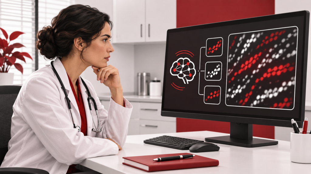
Artificial intelligence (AI) is being developed to detect suspicious regions, check whether anatomical areas have been adequately inspected, support photodocumentation and help characterize lesions. In upper GI endoscopy, AI may draw attention to a region that deserves a second look. In colonoscopy, computer-aided detection systems can flag lesions during withdrawal. AI tools for early GI cancer detection may be particularly useful as a second observer, helping clinicians maintain attention during a long examination.
However, an on-screen highlight is a prompt for assessment, not a diagnosis. Performance can vary with the device, population, lesion type and quality of the video. False alerts can distract, while a negative AI output cannot rule out disease. The ESGE position statement on AI sets performance expectations, including comparability with experienced endoscopists for key detection tasks. For gastric use, the 2025 ESGE guideline says real-time AI-assisted detection or localization may be used when available, while grading the supporting evidence as low quality.
For safe adoption, units should understand what their specific system has been validated to do, monitor false-positive and missed-lesion patterns, protect patient data and preserve clinician responsibility. AI can strengthen early GI cancer detection only when its limitations are visible and the human operator remains attentive to the mucosa, the patient and the whole clinical context.
Limitations, training and questions clinicians ask
Advanced imaging cannot compensate for poor preparation, inadequate visibility, incomplete examination or lack of operator experience. Equipment also differs between centres, and cost or workflow may limit access. A suspicious appearance may still need histological confirmation, while a normal-looking field cannot remove the need for appropriate surveillance in a patient with a known risk condition. Early GI cancer detection is therefore a combination of patient selection, high-quality technique, appropriate technology and reliable follow-up.
Training matters because optical patterns are learned, not self-explanatory. ESGE recommends structured learning based on validated classifications and continued practice to maintain competence. Teams can review representative cases, compare endoscopic impressions with pathology, discuss missed lesions and audit documentation. This approach encourages improvement without treating one detection rate or one AI tool as a complete measure of care.
Does advanced imaging replace biopsy?
No. It can help identify and characterize suspicious tissue, but biopsy or resection pathology remains important when required to establish diagnosis and guide treatment.
Is NBI used for every endoscopy?
No. NBI and other enhanced modes are selected according to the indication, equipment, lesion and endoscopist’s training. High-quality white-light inspection remains essential.
Can AI confirm that there is no cancer?
No. AI can assist detection in validated settings, but it cannot rule out cancer or replace a complete examination and clinical judgement.
Does everyone need an endoscopy for cancer screening?
No. Screening strategies differ by cancer type, individual risk and local recommendations. Endoscopy should be considered in the appropriate clinical context, not assumed to be necessary for everyone.
The key message is simple: advanced endoscopic imaging improves the opportunity to see and describe subtle changes, but good outcomes depend on the complete care pathway. Use validated tools, know their limits, document findings and make sure the patient has an appropriate plan.
Related SGEI resources:
- SITAR: SGEI School of Innovation, Training and Research training, research and academic initiatives.
- Young Endoscopist UpSkilling (YES) programme information and eligibility for early-career endoscopists.
- Journal of Digestive Endoscopy research, reviews and developments in digestive endoscopy.
- SGEI Events check the official page for current conferences and learning activities.
- Colonoscopy quality indicators a companion SGEI article on endoscopy quality measurement.
Join SGEI membership

Improving early GI cancer detection depends on continued learning, careful practice and collaboration across the endoscopy community. The Society of Gastrointestinal Endoscopy of India (SGEI) brings together professionals interested in advancing GI endoscopy through education, research and shared academic activity. Membership can help clinicians stay connected with resources, learning opportunities and peers working in the field.
If you want to take part in this community, explore the SGEI membership page for current information about membership, learning and collaboration. Review the latest categories and requirements, then use the SGEI membership application form if you are ready to apply. Eligibility, documents and fees may change, so confirm the current details on the official site before submitting an application.
Join SGEI and be part of a community shaping the future of gastrointestinal endoscopy in India.